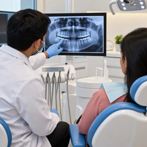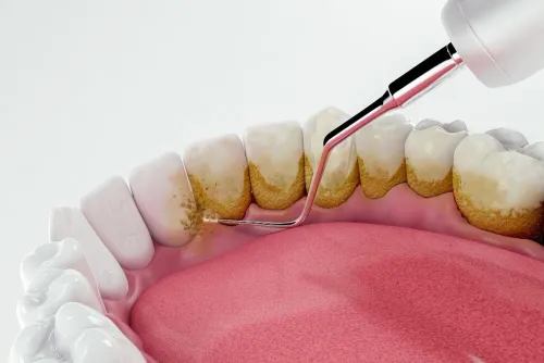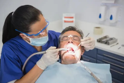
Many dental problems begin beneath the surface, where they cannot be seen during a routine oral examination. Cavities between teeth, hidden infections, bone loss, impacted teeth, and developing dental conditions often require imaging for accurate diagnosis. Professional Dental X-Ray Services play an important role in helping dentists detect these issues early and create precise treatment plans.
Modern dental X-rays use advanced digital technology to produce detailed images while exposing patients to significantly lower levels of radiation than older conventional systems.
Why Are Dental X-Rays Necessary?
A visual examination alone cannot reveal every dental problem.
Dental X-rays help dentists identify:
- Tooth decay between teeth
- Bone loss around teeth
- Dental abscesses
- Impacted wisdom teeth
- Root infections
- Jawbone abnormalities
- Development of permanent teeth in children
Early detection allows treatment to begin before conditions become more complex.
Different Types of Dental X-Rays
Dentists use different types of X-rays depending on the patient's needs.
Common imaging includes:
Bitewing X-Rays
Used to detect cavities between teeth and monitor existing fillings.
Periapical X-Rays
Provide detailed images of an individual tooth from crown to root.
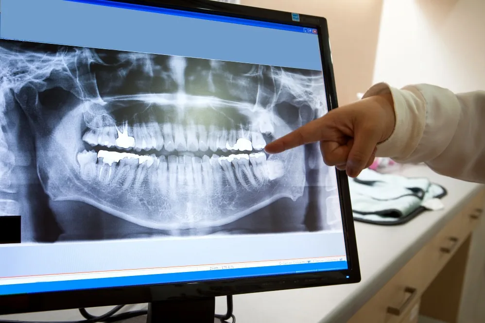
Panoramic X-Rays
Capture the entire mouth, including both jaws, wisdom teeth, and surrounding structures.
Occlusal X-Rays
Often used to evaluate tooth development and certain jaw conditions.
Your dentist recommends the appropriate imaging based on your symptoms and treatment requirements.
Are Dental X-Rays Safe?
One of the most common concerns patients have is radiation exposure.
Modern digital dental X-rays are designed to use very low radiation doses while producing high-quality diagnostic images. Additional protective measures, such as lead aprons and thyroid collars where appropriate, further enhance patient safety.
Dentists only recommend X-rays when they are clinically necessary.
How Dental X-Rays Support Better Treatment
Accurate diagnosis leads to more effective dental care.
Dental imaging assists with:
- Root canal treatment planning
- Dental implant assessment
- Orthodontic evaluations
- Wisdom tooth assessment
- Gum disease diagnosis
- Monitoring healing after procedures
Without proper imaging, some dental conditions may remain undetected until they become more advanced.
How Often Are Dental X-Rays Needed?
There is no fixed schedule for everyone.
The frequency depends on factors such as:
- Age
- Medical history
- Oral health status
- Risk of tooth decay
- Previous dental treatment
Your dentist determines when new imaging is appropriate based on your individual needs rather than following a routine timetable.
Preparing for a Dental X-Ray
Dental X-rays require very little preparation.
During your appointment:
- Remove metal jewellery around the head and neck if requested.
- Follow the technician's positioning instructions.
- Remain still while the image is captured.
- Inform your dentist about any relevant medical conditions.
The entire process is usually completed within a few minutes.
Why Digital Imaging Benefits Patients
Patients throughout Bengaluru increasingly benefit from digital dental imaging because it provides faster results, clearer images, and improved treatment planning. Immediate image availability also allows dentists to explain findings more effectively, helping patients better understand their oral health.
Digital technology supports earlier diagnosis and contributes to more predictable treatment outcomes.
The Value of Accurate Diagnosis
Successful dental treatment begins with understanding the exact cause of the problem. Atlas Dental Clinic uses modern digital dental X-ray technology to support comprehensive examinations and individualized treatment planning. By combining advanced imaging with clinical expertise, the clinic helps patients receive accurate diagnoses, timely treatment, and informed guidance for maintaining long-term oral health.
Keywords