Tooth pain, swelling or a suspected cavity does not automatically require the same examination or X-ray. Knowing what happens at each stage helps you prepare useful information, understand why an image is recommended and take part in the treatment decision that follows.
Key takeaways
- Bring your medical history, medicines, previous X-rays and a clear reason for visiting.
- Your symptoms and examination determine whether you need bitewing, periapical or panoramic imaging.
- Digital X-rays use a sensor, take seconds and require protective positioning before exposure.
- Ask the dentist what the image shows, what needs treatment and when follow-up is needed.
What happens when the dental consultation begins?
Registration confirms your identity, contact details and reason for visiting; the dentist then turns that information into a clinical assessment rather than starting with an X-ray.
1. The dentist reviews your dental and medical history, including previous treatment, current medicines, allergies and conditions that affect healing or bleeding. Explain the tooth, gum or jaw problem in your own words, including when it began, what triggers it and whether you have pain, swelling, sensitivity or difficulty chewing.
2. An extraoral examination checks your face, jaw movement and any visible swelling or asymmetry. The dentist may feel the jaw joints and nearby tissues if you report clicking, restricted opening or facial pain.
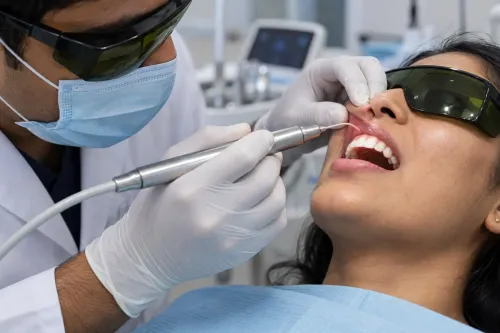
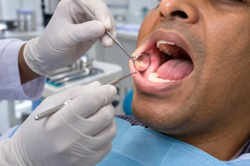
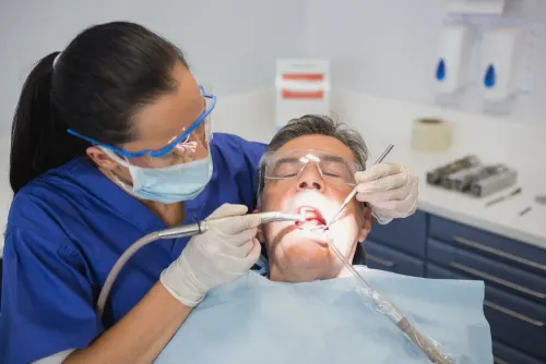
3. During the intraoral examination, the dentist uses a mirror, light and probe to inspect each tooth, existing fillings or crowns, the gums, tongue, cheeks, palate and other oral tissues. They look for decay, broken restorations, inflammation, bleeding, pockets and signs of infection.
4. The dentist checks your bite by watching how your upper and lower teeth meet and asking you to open, close and move your jaw. This helps identify uneven contact, tooth wear, clenching patterns or a bite problem affecting comfort and function.
The findings guide the next discussion. You may receive an immediate diagnosis, advice to monitor a change, or a recommendation for imaging or another test when the examination alone cannot answer the clinical question.
What should you bring and explain before the examination?
Bring your medication list, medical history, previous dental records and a clear description of your symptoms. This information gives the dentist a safer starting point for the dental consultation process and helps distinguish a tooth problem from a wider health issue.
- List every prescription, over-the-counter medicine, supplement and blood thinner, including its dose.
- Name allergies to medicines, latex or other materials, and describe the reaction.
- Report diabetes, heart disease, bleeding disorders, immune conditions, osteoporosis treatment, recent surgery and any major illness.
- State whether you are pregnant or might be pregnant before any X-ray is considered.
- Bring previous X-rays, treatment notes, implant or crown details, and a list of recent dental procedures.
- Explain when the problem began, what triggers it, whether it is worsening, and whether pain is sharp, throbbing, constant or temperature-sensitive.
- Point out swelling, pus, bleeding, bad taste, fever, numbness, difficulty opening your mouth, chewing or swallowing.
| Information | Tell or bring | Why it matters |
|---|---|---|
| Pain | Tooth location, severity, duration and triggers | Helps identify possible decay, crack, gum disease or pulp irritation |
| Swelling | Location, onset, drainage and fever | Signals infection risk and urgency |
| Previous treatment | Fillings, root canals, extractions and trauma | Prevents misreading altered teeth or recurring problems |
| Medical history | Conditions, medicines and allergies | Changes examination, imaging and treatment safety |
If you cannot remember a medicine’s name, bring its packet or a photograph of the label.
Why might the dentist recommend one type of X-ray rather than another?
Imaging is recommended when your symptoms, history and examination leave a question that an image can answer. A dentist first checks your teeth, gums, restorations, bite and jaw function; if the findings are clear and no hidden problem is suspected, an X-ray may not be needed.
This is how a digital dental diagnosis avoids treating the image as the diagnosis.
The choice depends on the area and detail required:
| Type | What it shows | When it applies |
|---|---|---|
| Intraoral | One or a few teeth in close detail | Suspected decay, cracks, bone loss or root changes |
| Bitewing | Upper and lower back teeth and the bone level between them | Detecting cavities between teeth and assessing crestal bone |
| Periapical | The full tooth, root tip and surrounding bone | Toothache, infection, abscess, trauma or root-canal assessment |
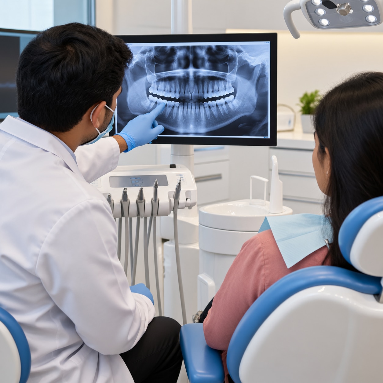
| Panoramic | Both jaws, all teeth and nearby structures in one broad image | Impacted teeth, jaw lesions, orthodontic review or extensive planning |
| CBCT | A three-dimensional view of teeth, roots, bone and anatomical structures | Selected implant, surgical, orthodontic or complex endodontic planning |
A panoramic image is broad but less detailed because structures overlap, so it does not replace a targeted bitewing or periapical image. CBCT is not simply a sharper routine X-ray; its generally higher radiation dose makes it unsuitable when examination or two-dimensional imaging can answer the question.
The dentist aims to obtain the necessary information with the lowest reasonable exposure. “Digital” does not mean radiation-free, and movement or poor positioning can require a repeat image.
What happens before, during and after a digital X-ray appointment?
A dental X-ray appointment in Bangalore usually takes only a few minutes, but the image depends on accurate positioning. The radiographer selects the view needed, explains where to place your teeth or head, and confirms pregnancy or possible pregnancy before exposure.
1. You may bite gently on a holder, or the radiographer may place a small electronic sensor inside your mouth. For a panoramic image, you stand or sit with your chin and forehead supported while you keep your tongue in position.
2. Stay still and follow the breathing or biting instruction during the brief exposure. Movement, incorrect angulation or a sensor covering the wrong area can blur the image or hide a root, leading to a repeat.
3. The dentist or radiographer checks the image for coverage, sharpness and exposure before you leave. Digital images appear quickly and can be enlarged or adjusted for viewing, but a clear image does not replace the clinical examination.
4. Radiation protection means taking only the views needed, using correct exposure settings and beam restriction, maintaining the equipment and avoiding unnecessary retakes. Digital does not mean radiation-free. Tell the team if you are pregnant or might be pregnant so they can weigh urgency, alternatives and local safety protocol.
If you are comparing a digital x ray dentist kammanahalli with another provider, ask how image quality is checked and how repeats are handled. At Atlas Dental Clinic, use the review to ask which finding the image supports and which symptoms still require examination.
Related service
Consultation and Digital X-ray Consultation and Digital X-ray At Atlas Dental Clinic, located in Kammanahalli, Bangalore, we provide comprehensive dental consultation and digital X-ray services to accurately... View service → |
What can the image show, and what happens after the results are reviewed?
An X-ray can show cavities between teeth, bone loss, a root fracture, an abscess, or changes around a tooth root. A periapical image shows the whole tooth from crown to root tip, helping investigate root-surface disease, trauma and unusual pain.
During a dental consultation and digital X-ray in Kammanahalli, the image is one part of the assessment. A normal result does not exclude early decay, cracks, pulp inflammation or soft-tissue disease. Symptoms, examination, cold or tapping tests, and comparison with earlier images can change the interpretation.
Digital dental diagnosis should end with a decision, not an automatic filling, extraction or root canal. Ask the dentist to identify each finding, explain its severity, and compare treatment, prevention, monitoring or further tests such as CBCT or a repeat radiograph. Discuss benefits, risks, stages and costs, and ask what change would trigger treatment.
Frequently asked questions
What happens during a dental consultation?
Registration confirms your details and reason for visiting. The dentist reviews your history, asks about symptoms, examines your mouth and recommends tests or treatment.
What should I bring to a dental consultation?
Bring previous dental records or X-rays, a list of medicines, relevant medical conditions, allergy information and details about pain, sensitivity or swelling.
Why does a dentist recommend different types of X-rays?
Each image covers different structures. Bitewing X-rays show spaces between back teeth, periapical X-rays show an entire tooth and root, and panoramic X-rays show both jaws.
What happens during a digital dental X-ray appointment?
The technician positions a sensor or imaging device, asks you to stay still and captures the image in seconds. The dentist then reviews it with your examination findings.
What can a digital dental X-ray show?
It can reveal cavities between teeth, bone loss, infections near roots, impacted teeth, dental work problems and changes that cannot be seen during a visual examination.
Keywords